Public Data Sets: GroEL I
1. Images
Images were acquired with a FEI Tecnai F20 equipped with a 2Kx2K CCD Tietz camera, as defocal pairs at a nominal magnification of 62,000 x and a voltage of 120 KeV, using the Leginon system (Potter et al., 1999 ; Carragher et al., 2000). The first image (named *.001.mrc) is acquired at near to focus (NTF) conditions (-0.6 to -1.5 µm) and the second image (named *.002.mrc) at farther from focus (FFF) conditions (-3µm). The time interval between the two exposures is approximately 20s due to the time required to read out the digital image from the camera. The pixel size is 2.238 Å at the specimen scale and the accumulated dose for each high magnification image area was ~10 e/Ų.
Below is an example of a defocus pair. Click on the images to see at full scale.
(a)  (b)
(b) 
(a) Near to focus (NTF) image. (b) Far from focus - FFF image.
Figure 1: An example pair of high magnification images of GroEL.
Downloading High Magnification Images: Image files are provided in MRC format and as JPEG files
Download entire set of images here (~52GB).
We suggest using a tool called em2em, to convert MRC files to other formats.
2. Positions of the Picked Particles in the Images
Particles are picked in the FFF images and these coordinates are used to select particle in the NTF images. Phase correlation is used to align the two images to calculate the offset between the FFF coordinates and the NTF coordinates.
(a)  (b)
(b) 
Figure 2: Screenshots of using the wizard with the above tips to download the whole set of high magnification images.
2.1 Automated Picking:
A two-stage approach was applied for automatic selection of GroEL particles. In the first stage, a set of candidate particles were automatically selected for each micrograph using the fast local correlation algorithm (Roseman, 2003) combined with a peak searching and thresholding algorithms. In the second stage, the set of candidate particles were filtered with a clutter finder to reject contamination (Zhu et al., 2004). The clutter finder starts with edge detection followed by connected component labeling, convex hull computation, and size filtering. Particle coordinates are provide below:
Coordinates of 16212 side-view and 10603 top-view GroEL particles selected in the FFF images. [Need a description of the coordinate file – the link is missing in the pages at the moment]
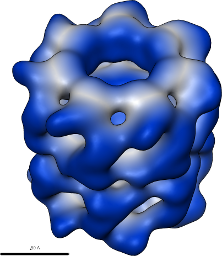
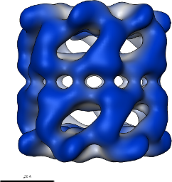
3. 3D Reconstructed Map
A set of 7505 particles were extracted from a set of 141 NTF micrographs whose corresponding FFF micrographs contain the first 7,505 particles of the automatically selected 16216 side-view particles. This particle set was used to create a 3D reconstruction using D7 point-group symmetry and 9 cycles of refinement.
3D map in MRC format of 7,505 side-view particles contributing to the map.
Table 2: Sample reconstruction using 7,505 side-view particles.
Updated by Eric Hou almost 15 years ago · 31 revisions